Today was my first day at the National Toxicology Program with Dr. Malarkey inside the National Institute of Environmental Health Science site. First I had to go through security clearance at the front gate, showing my ID and my incredible smile (they knew I wasn’t a criminal). We then drove through the campus to reach building 101, which was a huge building overlooking their lake. I received my visitor ID and met Anthony in the front lounge who was checking in at the same time. We waited in the front couches until Dr. Malarkey came to pick us up. We went up to the third floor of the building, and Dr. Malarkey showed me the cubicle Anthony and I would be sharing. There were already a few abstracts on the countertop that I would need to read, to familiarize myself with the cell phone radio frequency radiation study. We made the short walk from our cubicle to his office, where he showed us his pathology museum. This consisted of shelves full of antiques and artefacts including his first set of miniature microscopes and slides, horse intestine stone, emu egg, and old books. We looked at a few of the old slides through the two person microscope he had in his office, looking at lobster larvae.
We set Anthony up with a few slides of the eye and Dr. Malarkey asked him to identify the different parts in the eye, location and type of tumor, and type of tissue and cells. While Anthony was squinting into the microscope, Dr. Malarkey gave me a tour of the third floor office and an introduction to what he and the pathology group does. In his doctorate program at NC State, Dr. Malarkey specialized in looking at liver cancer in mice. Hence, he went to teach in a lot of different countries after becoming well-known for this research. His research and focus still involves testing rat and mice, but the most important study of present is looking at the effects of cell phone radio frequency radiation on the presence of cancer. The National toxicology program also releases thick reports every few months about the results of the research they have been conducting. Dr. Malarkey explained how these thorough documents take close to ten years to prepare with almost 30 people working on each edition. We looked at a few together, all looking at how different chemicals, radioactive sources, or drugs effect the chance for cancer. To do this they test the materials on mice, rats, fish, and genetically modified mice. Most studies use mice and rats as the group, testing both species male and female.
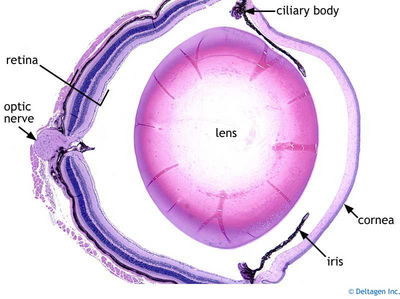
After this, we all went downstairs to the ten person microscope to look at the slides Dr. Malarkey had given Anthony. After focusing all of our individual eyepieces, we determined that the first slide was actually a cross section of an eye. Dr. Malarkey helped us to name and locate each part of the eye including the optic nerve, lens, cornea, etc. The slide also had a tumor on the right and left sides of the optic nerve, and helped us characterized each type of nerve so that we could recognize them in this slide and the next ones. We then looked at other slides, including one with a cat who had cataracts. After discuss more characterizations of cancer, cells, and learning that blue means nucleus (bad) and red means blood, we headed to lunch.
After lunch, Anthony and I went to the histology labs to watch how samples and slides are made. First we visited the Necropsy department to watch the dissection of a mouse. The mouse was killed with carbon monoxide poisoning, and then the scientist covered it’s skin in ethanol. Using scissors the scientist cut open the mouse’s skin, and carefully removed all organs in the mouse. With our careful observation, we were then allowed to dissect a mouse of our own, removing every organ as well except for the esophagus.
I had so much fun today and am so excited for tomorrow!